

Importance of Animal Health
( पशुओमेंस्वास्थ्यकामहत्व )
The animal, a cow or buffalo that produces milk should be free from zoonotic diseases and other diseases of Veterinary importance, so that the quality and quantity of milk will be enriched automatically. What if a cow producing 10-20 Litres of Milk get diseased treated with antibiotic and needed to be under withdrawl period of milk, even if not followed keeping in mind about the quality and quantity loss of milk, the economic loss incurred to the farmer will be high and intangible. These losses are generally inaccountable and inevitable.

More Info
Diseases in Animals
There are many diseases in milch animals due to various reasons. Micro viruses, bacteria, fungi, parasites, protozoa, protozoa, malnutrition and disorders in the metabolism inside the body are among the main reasons. There are many life-threatening diseases in these diseases, many diseases adversely affect the production of animals. Some diseases are transmitted from one animal to another, such as mouth and hoof disease, strangulation, etc., are called contagious diseases. Some diseases also come from animals to humans, such as rabies, tuberculosis, etc., these are called zoonotic diseases. Therefore, it is necessary for the animal husband to be aware of the major diseases so that by taking appropriate steps at the right time, he can protect himself from financial loss and also cooperate in protecting human health. Following are the major diseases of milch animals:
More Info
Cowpox - smallpox disease
Cowpox - Chickenpox Cowpox is an infectious disease caused by the cowpox virus. The virus, part of the genus Orthopoxvirus, is closely related to Vaccinia virus. The virus is zoonotic, meaning it is transferable between species, such as from cat to human. Transmission of the disease was first observed in dairy maids who touched the udders of infected cows and consequently developed the signature pustules on their hands. Smallpox is more common in animals other than bovines, such as rodents. Smallpox is similar to the highly contagious and often fatal smallpox disease, but is much milder than that. Its proximity to the milder form of smallpox and the observation that dairy farmers were immune to smallpox inspired the modern smallpox vaccine, which the English physician Edward Created and administered by Jenner. The term "vaccination", coined by Jenner in 1796, is derived from the Latin adjective vaccinus, meaning "of or from a cow". Once vaccinated, a patient develops antibodies that immunize them to smallpox. but they also develop immunity to the smallpox virus, or variola virus. Smallpox vaccination and subsequent incarnations proved so successful that in 1980, the World Health Organization declared that smallpox was the first disease to be eradicated by worldwide vaccination efforts. [4] Other orthopox viruses are prevalent in some communities and continue to infect humans, such as cowpox virus (CPXV) in Europe, vaccinia in Brazil, and monkeypox virus in Central and West Africa.

Medical use
Naturally occurring cases of smallpox were not common, but it was discovered that the vaccine could be "carrying" in humans and reproduced and transmitted from human-to-human. Jenner's original vaccination used lymph from a smallpox blister on a milkmaid, and later "hand-to-hand" vaccinations applied the same principle. Since this transfer of human fluids came with its own set of complications, a safer method of producing the vaccine was first introduced in Italy. The new method used cows to manufacture the vaccine using a process called "retrovaccination", in which a heifer was infected with a humanized smallpox virus, and it was passed from calf to calf to grow efficiently and safely. was passed to produce in quantity. This was followed by the next incarnation, the "true animal vaccine", which used the same process but started with the naturally occurring smallpox virus, not the humanized form. This method of production proved lucrative and was taken advantage of by many entrepreneurs requiring only calves and seed lymph from an infected cow to manufacture crude versions of the vaccine. National Vaccine Establishment's W.F. Elgin presented his slightly refined technique at a conference of the State and Provincial Boards of Health for North America. A tuberculosis-free calf, abdomen shaved, will be tied to an operating table, where incisions will be made on its lower body. Glycerin-containing lymph from a previously vaccinated calf was spread along the cut. After a few days, the cut may have scratched or crusted over it. The crust was softened with sterilized water and mixed with glycerin, which disinfected it, then hermetically sealed in capillary tubes for later use. At some point, the virus in use was no longer smallpox, but vaccinia. Scientists have not determined exactly when the change or mutation occurred, but the effects of vaccinia and cowpox virus as vaccines are almost identical. [5] The virus is found in Europe and mainly in the UK. Human cases are very rare today and are most often contracted from domestic cats. The virus is not usually found in cattle; Reservoir hosts for the virus are woodland rodents, particularly voles. From these rodents, domestic cats contract and transmit the virus to humans. [6] Symptoms in cats include sores on the face, neck, forearms and paws, and less commonly, upper respiratory tract infections. [7] Symptoms of infection with chickenpox virus in humans are localized, usually with pustular lesions on the hands and limited to the site of introduction. The incubation period is 9 to 10 days.
Search
In the years 1770 to 1790, at least six people who came into contact with cows independently tested the possibility of using the smallpox vaccine as a form of immunization for smallpox in humans. Among them were the English farmer Benjamin Zesty in Dorset in 1774 and the German teacher Peter Pellet in 1791. Zesty vaccinated his wife and two young sons with smallpox, in a successful attempt to immunize them from smallpox, an epidemic of which had originated in their town. His patients who had contracted and recovered from similar but mild chickenpox (mainly milder) were immunized not only for further cases of smallpox, but also for smallpox. By scraping the fluid into the skin of healthy individuals from smallpox wounds, he was able to immunize those people against smallpox. Reportedly, farmers and those who regularly worked with cattle and horses during smallpox outbreaks were often spared. An investigation by the British Army in 1790 revealed.
Bovine Abortions
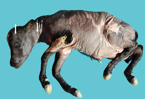
Bovine Abortions - - Bovine abortion Bovine abortion of unknown infectious pathology still remains a major economic problem. In this study, we focused on a new potential abortive agent called Parachlamydia acanthamoebae. Retrospective samples (n = 235) taken from late gestation in cattle were screened for Chlamydiaceae and Parachlamydia spp., respectively, by real-time diagnostic PCR. Histological sections of positive cases were further examined by immunohistochemistry using specific antibodies by real-time PCR for any chlamydia-related agent. Chlamydophila abortion was detected by real-time PCR and arraytube microarray in only three cases (1.3%) playing a less significant role in bovine abortion than in the case of small ruminants in Switzerland. Of the 235 (18.3%) cases, 43 turned out to be positive for parachlamydia by real-time PCR. The presence of parachlamydia within placental lesions was confirmed by immunohistochemistry in 35 cases (81.4%). The main histopathological feature in parachlamydial abortion was purulent to necrotizing placentitis (25/43). Parachlamydia should be considered as a new abortive agent in Swiss cattle. Because Parachlamydia may be involved in lower respiratory tract infections in humans, bovine abortion material should be handled with care given the potential zoonotic risk.
Summary
Neospora caninum is an apicomplexan protozoan that causes abortion in cattle worldwide. The present study was designed to assess the importance of bovine neosporosis in causing abortion in Iranian cattle. Infection was primarily diagnosed by polymerase chain reaction (PCR), complemented by histopathology and immunohistochemistry (IHC). One hundred brains of aborted bovine embryos were collected from an Iranian dairy herd in the Mashhad region between 2003 and 2005. in the brains of 13 aborted fetuses by PCR. caninum was detected. In 12 of the fetal brains, N. caninum infection were observed. Immunohistochemical examination of the brain revealed N. caninum organisms, and a thick-walled (2 µm) cyst with a diameter of 50 µm was identified in one of the IHC-positive brain. The results indicated that neosporosis is an important cause of abortion in dairy cattle of Iran.
Materials and methods
Relevant articles were searched in February 2016 using four relevant scientific databases (PubMed, Web of Science, CAB Abstracts, and Agricola)
Result
Initial database searches yielded a total of 1724 articles that matched the search terms. After removing citations and studies irrelevant to the current meta-analysis (ie, that did not evaluate the efficacy of BoHV-1 vaccination in abortion prevention), 41 full-text articles were evaluated for study merit. A total of 15 studies in 10 manuscripts were identified for inclusion, after the exclusion of 26 reports for incompatibility with study objectives or design.
Discussion and conclusion
This study provides cumulative and quantitative evidence of the efficacy of BoHV-1 vaccination in reducing the risk of miscarriage in pregnant cattle. A meta-analysis of controlled studies examining the efficacy of vaccine protection demonstrated a 60% reduction in the risk of miscarriage in vaccinated cattle. This study confirms the benefit of using vaccination as a component of an overall herd health program to reduce the negative effects of BoHV-1 infection.
Leptospirosis
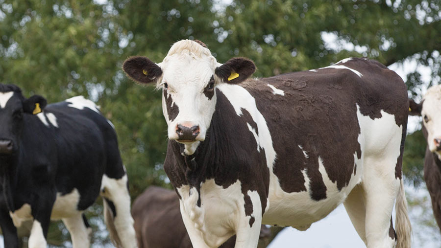
Leptospirosis - is a common infection in dairy and beef herds that causes infertility, miscarriage and poor milk yield. Resulting in infertility, miscarriage, poor milk yield. Leptospirosis affects humans causing influenza-like symptoms with severe headache but can be treated effectively. Dairy farmers are especially at risk of infection due to urine splashing on their faces while feeding cows. Pasteurization destroys all the leptospore organisms excreted in the milk.
Leptospirosis cause
Leptospirosis is a zoonotic disease caused by bacteria of the genus Leptospira. Different serogroups are often more prevalent depending on the location. Some examples of different serovars include hardjaw, pomona, canicola, icterohemorrhagia, and gripotyphosa. Cattle are the maintenance host for hardjaw, but since it is specialized to survive within cattle, the infection is less severe. Animals infected with other strains (such as Pomona) suffer from more severe disease. Maintenance hosts carry the bacteria and expose other susceptible animals. Maintenance hosts may be cattle, pigs, dogs, rodents or horses. An animal can become infected with serovars maintained by its own species (maintenance host infection or host-adapted infection) or serovar created by other species (accidental infection or non-host-adapted infection). Leptospirosis is transmitted either directly between animals or indirectly through the environment.
Symptom
Clinical signs of lepto depend on the degree of resistance or immunity of the herd, the infected serovar and the age of the infected animal.
Host-optimized
Leptospira hardjo-bovis is the only host-adapted lepto serovar in cattle and can infect animals at any age, including young calves. Because cattle are maintenance hosts for Hardjo-bovis, infection with this serovar will often produce a carrier state in the kidney associated with prolonged urinary effusion. In addition, infection with Hardjo-bovis can persist in the reproductive tract. Infertility that can result from frequent reproductive tract infections is perhaps the most economically damaging aspect of leptospirosis. Low antibody titers are commonly associated with Hardjo-bovis infection, making it difficult to detect and diagnose.
Non-Host-optimized
Lepto serovars include Leptospira pomona, Icterohemorrhagia, Canicola and Gripotyphosa. As cattle are the casual hosts for these lepto serovars, clinical symptoms are usually very different from those of Hardjo-bovis infection. When leptospirosis associated with non-host-adapted lepto serovar occurs in calves, the result is high fever, anemia, red urine, jaundice, and sometimes death in three to five days. In older cattle, early symptoms such as fever and lethargy are often mild and usually go unnoticed. In addition, large animals usually do not die from leptospirosis. Lactating cows produce less milk, and the milk they give for a week or more is thick and yellow. Leptospirosis with non-host-adapted lepto serovar also affects pregnant cows causing fetal death, miscarriage, stillbirth, retained placenta and birth of weak calves. Miscarriage usually occurs three to ten weeks after infection.
Treatment
Antibiotic therapy should be prescribed for animals with leptospirosis. Antibiotics can also eliminate persistent infections. Infected animals should be isolated from others to avoid transmission of the disease. The treatment has been shown to be most effective when animals are treated during periods of leptospiremia. However, antibiotic therapy during chronic infection may reduce carrier status. When there is a storm of infection through the herd, especially when multiple pregnant cows are involved, simultaneous treatment and vaccination of all animals will reduce new cases and miscarriages if the treatment is administered early in the herd's infection.
Redressal
Vaccination is relied upon to increase immunity to infection. The primary course of vaccination consists of two injections four weeks apart and is followed by an annual increase. Vaccination should prevent urine flow after exposure and prevent weaning and miscarriage. Annual vaccination should be used in closed herds, while semiannual vaccination should be considered for open herds. Calves born from vaccinated cows are only immunized for six months, and will need their own schedule of vaccinations. Management methods to reduce transmission include rat control, fencing of cattle from potentially contaminated streams and ponds, isolating cattle from pigs and wildlife, selecting replacement stock from herds seronegative for leptospirosis, and chemoprophylaxis and replacement stock. Vaccination is included. In some cases, streptomycin is added to the semen of bulls kept at artificial insemination centers as a precaution.
Metabolic Disorders
Metabolic disease refers to a group of conditions caused by a deficiency of certain essential nutrients that result in disturbances in the normal metabolic processes of the animal. These conditions are multifactorial and usually occur at times of high physical stress or demand for these nutrients with late pregnancy and early lactation. Signs of these conditions may overlap and look similar and it is not uncommon for more than one disease to occur at the same time, further complicating the picture. Therefore, it is important to understand the causes of these diseases as prevention and treatment are different.
Redressal
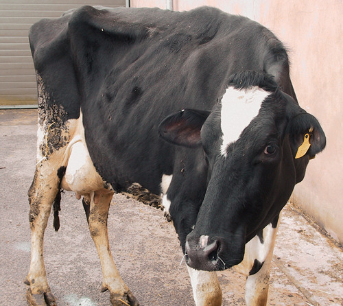
These are diseases of livestock caused by productivity practices when the body stores on calcium, magnesium or energy that cannot meet metabolic needs. They are very important in places where high production animals are required, e.g. in the diary industry. In cattle, metabolic diseases include ketosis, milk fever, fat cow syndrome and hypomagnesemia. All of these can produce an acute, temporary, but potentially fatal deficiency. Improving the diet of cows during the period from late pregnancy to peak lactation is important to prevent these diseases. If these diseases recur, it is necessary to seek professional veterinary and nutritional advice.
Summary
Improved reproductive performance and lower incidence of metabolic disorders have been considered as benefits of feeding supplemental fat to dairy cows. The increase in plasma non-esterified fatty acid concentrations during fat supplementation may be due to incomplete mobilization of fatty acids following lipoprotein lipase hydrolysis of very low-density lipoprotein triglyceride; However, evidence suggests that net adipose tissue triglyceride hydrolysis may be increased during fat supplementation. Plasma 3-OH-butyrate concentrations remain relatively constant during fat supplementation, but may have a tendency to decrease if fat is supplemented with cows with relatively high basal plasma 3-OH-butyrate concentrations. Since plasma ketone levels typically increase when non-esterified fatty acid concentrations are increased, it is hypothesized that the potential antiketogenic effects of excess fat are due to a glucose-sparing effect. The supplement does not appear to reduce hepatic lipid infiltration near the time of fat deposition. Possible mechanisms by which supplemental fat may improve reproductive performance include stimulation of prostaglandin F2 A synthesis and secretion and enhanced utilization of blood cholesterol for progesterone synthesis. The days leading up to the first ovulation and the luteal function of dairy cattle have been correlated with energy balance during the first 3 weeks postpartum. Energy balance data for early-fed cows with supplemental fat are not plentiful; However, a slight but statistically insignificant increase has been observed when feeding fat. Cows that are fed supplemental fats experience better energy balance, with increased follicular growth and development being able to start the cycle sooner. Applied studies examining the effects of supplemental fat on reproductive performance have provided inconsistent results.
Mouth-pocket
Foot-and-mouth disease, FMD or hoof-and-mouth disease is a highly contagious and fatal viral disease of split-hoofed animals. This happens to domestic animals like cows, buffaloes, sheep, goats, pigs and wild animals like deer etc.
Reason
This disease is spread through contact, contaminated water and feed. This disease is caused to animals by a very small worm that cannot be seen by the eye. Which is called virus or virus. Mouth-foot disease can occur in cows of any age and their babies. No season is fixed for this, that is to say, this disease can spread in the village at any time. Although cows do not die from this disease, yet milch animals dry up. This disease is sometimes fatal for adult animals, but milk production and reproductive capacity in cows and bearing capacity in bullocks suffer even after health benefits. It is generally fatal in calves and calves. It also affects sheep, goats and pigs. Since there is no cure for this disease, it is beneficial to get vaccinated before the disease occurs.
Symptom
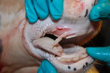
When this disease comes, the animal gets high fever. In the mouth, gums, and the inner part of the lips above the tongue of the sick animal, small grains emerge in the space between the hooves, then gradually these grains join together to form a big blister. Over time, these blisters become fruitful and they get injured. In such a situation the animal stops chewing. All the saliva falls from the mouth. Animals become lethargic. Doesn't eat or drink anything. Due to the wound in the hoof, the animal walks with a limp. When mud, soil etc. is applied to the wounds of the feet, then insects fall in them and they feel very painful. The animal starts limping. Milk production in milch animals drops completely. They start getting weak. These blisters and wounds get filled with time and treatment, but in hybrid animals this disease can sometimes cause death. It is a viral disease that affects animals with cloven foot. Generally cattle of cow, buffalo, sheep, goat and pig species come under its grip. It is a contagious disease.
Main characteristics
Swelling of the foot (around the hoof) Lameness of the foot that is short-lived (one to two days) Fever in the hoof and maggots in the wounds Sometimes from the foot of the hoof Separation, drooling of mouth, blisters appear on the tongue, gums, lips, etc., which later break down, resulting in excessive decline in production capacity, decrease in the working capacity of the bulls, the affected animal continues to gasp for months even after being healthy. Even after recovery, the fertility of animals remains affected for years. The fur and hooves of the body increase greatly, there is a possibility of miscarriage in pregnant animals.
The treatment
Vaccination of animals of 4 months or more age should be done once in 6 months, infected animals should be separated from healthy animals immediately because the secretions, dung and urine coming out of the body of infected animals contain virus. Food and green-dry fodder that came in contact with infected animals should be destroyed. All equipment used by infected animals should be sanitized and disinfected in 4% sodium carbonate solution or as prescribed by a veterinarian. The person who takes care of infected animals should be kept away from healthy animals. The infected area should be disinfected with 4% sodium carbonate solution or as prescribed by the veterinarian. The disease can be controlled by vaccination of sheep, goats and pigs. Immediate intimation to the concerned authority will help them to take necessary action for disease control which will help in controlling the spread or spread of the disease. The feet of the diseased animal should be washed two to three times a day by making a decoction of neem and peepal blisters. Wash the affected feet with phenyl-containing water two to three times a day and use fly repellent ointment. The blister should be washed thrice a day by dissolving 1% alum i.e. 1 gram alum in 100 ml water. During this time animals should be given soft and digestible food. Medication should be given on the advice of a veterinarian. Caution: The affected animal should be kept away from other healthy animals in a clean and ventilated place. The person taking care of animals should also come in contact with other animals after cleaning their hands and feet properly. The saliva falling from the mouth of the affected animal and the objects coming in contact with the foot wound should be burnt or the pit should be dug in the ground and buried with lime. Animals should be kept in a dry place so that our animals are safe.
Management
Its only symptomatic treatment is possible. Apply emollients to the wounds to reduce the pain. Consult a veterinarian for appropriate advice. Vaccination is better than cure On the principle of prevention, healthy animals above six months of age should be vaccinated against hoof-mouth disease.
Black Quarter Lame Fever
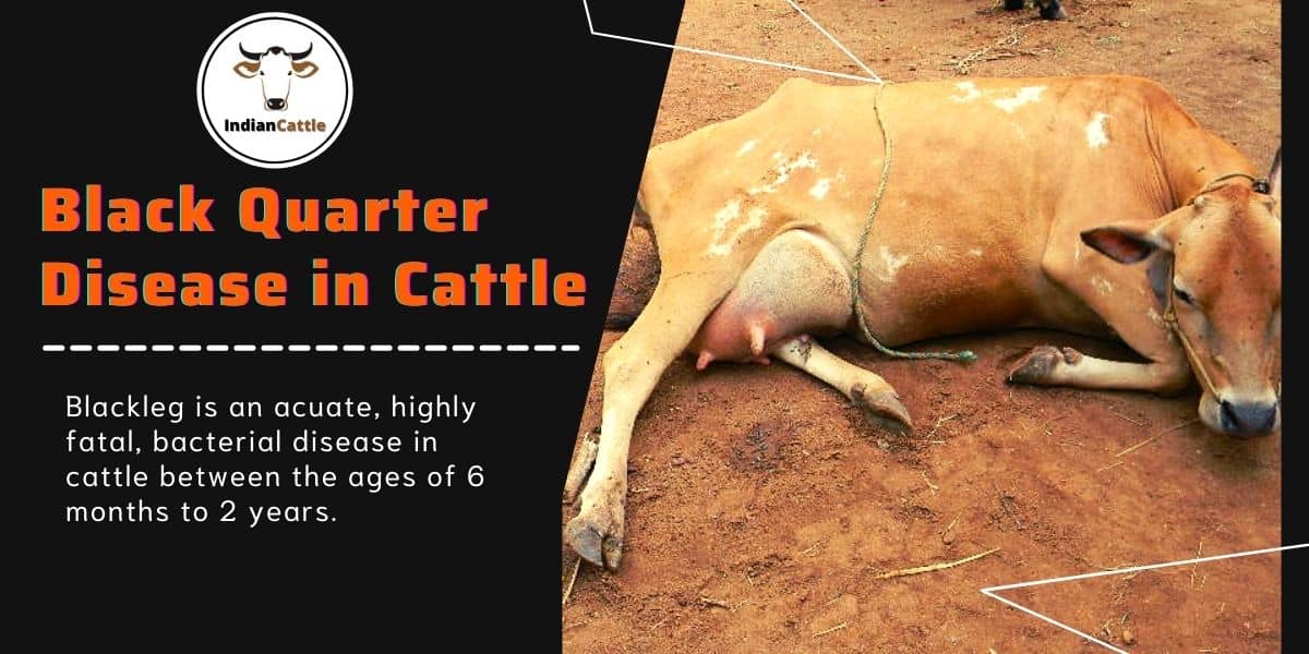
In common language, it is also known by names like Jaharbad, Phadsujan, Kala Bai, Krishnajandha, Langadiya, Ektanga etc. This disease is usually found everywhere but spreads widely in moist areas. Mainly, cow, buffalo and sheep are affected by this disease. Especially there is a bacterial disease of cows, in which swelling comes along with filling air in the muscles. Buffalo suffers very less from this disease, this disease is found more in animals between the age of six months to two years. This disease is famous by the name of 'Langdi disease'. It is an animal disease caused by bacteria (clostridium choviai bacteria), often occurring in cattle and occasionally in buffalo and sheep (Clostridium choviai bacteria), in which common fever and painful swelling of the fleshy part and lameness are the main symptoms. Young and healthy animals are more affected.
Symptoms of disease
This disease is more common in cattle. In this disease, the animal gets high fever and its temperature reaches from 106 degree F to 107 F. The animal becomes lethargic and stops drinking. There is heavy swelling in the upper part of the hind and fore legs of the animal. Due to which the animal starts walking with a limp or sits down. Due to the accumulation of gas, there is a crackling sound when the swollen area is pressed. The animal is unable to walk. This disease usually affects the hind legs more and swelling occurs in the area above the knee. This swelling is initially hot and painful, which later becomes cold and painless. Apart from the legs, swelling can also occur on the back, shoulders and other muscular parts. The skin over the swelling dries up and becomes tight. The treatment of the animal should be done soon because the poison caused by the bacteria of this disease spreads completely in the body and the animal dies. Procaine penicillin is very effective in this disease. Preventive vaccines are given for this disease. Mortality rate: 80-100 percent.
Prevention and prevention
This disease should be vaccinated before the rainy season. This vaccine is also given to the animal at the age of 6 months. Diseased animals should be separated from healthy animals. Vaccination should be done three months before shearing wool in sheep because the bacteria enter the body from the wound when the wool is sheared, which increases the chances of disease. The swelling should be opened by making an incision so that the bacteria become unaffected when exposed to air. Burning the upper layer of the soil with chaff in a particular area helps to eliminate the pathogens of the disease, at the time of pressing the dead body, the chosen pulse should be given on it.
The treatment
Treatment can be effective in the early stages of the infection, yet treatment is not beneficial in most cases. The use of antibiotics of penicillin, sulfonamide, tetracycline group along with supportive drugs is beneficial depending on the severity of the disease and the condition of the animal. Dressing with 2% hydrogen peroxide and potassium permanganate by making an incision in the inflamed area is beneficial.
Animal Epidemic or Rinderpest
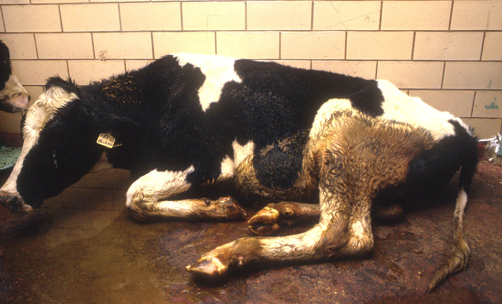
(Rinderpest) was a viral infectious disease caused by a viral filterable virus {Mensillus rinderpest distemper (MRD)} which has now been eliminated from the world. It was used by buffaloes and some other animals. The animal suffering from this disease used to see symptoms like fever, diarrhoea, drooling from the mouth, the sound of cuts from the teeth, blood (mucous) with cow dung. This disease is also a contagious disease caused by a virus that affects almost all ruminant animals. In this the animal gets severe diarrhea or dysentery. The disease is spread by direct contact of a healthy animal to a sick animal. In addition, this disease can also be spread through children and caregivers. In this the animal gets high fever and the animal becomes restless. Milk production is reduced and the eyes of the animal turn red. After 2-3 days, a rash appears in the mouth of the animal under the lips, gums and tongue, which later takes the form of a wound. The animal starts salivating from its mouth and gets thin and foul-smelling stools in which blood also starts coming. In this the animal becomes very weak and there is a shortage of water in it. In this disease the animal dies in 3-9 days. The outbreak of this disease used to kill millions of animals worldwide, but now under the Rinderpest Eradication Project implemented by the Government of India under the plan of eradication of this disease globally, now with the use of 100% preventive vaccines continuously. The disease has almost ended in the state and the country. A disease called 'rinderpest' had taken the form of an epidemic in the Middle East, Africa and Asia at one time. Due to this thousands of milch animals died. It is expected that after 'smallpox', 'rinderpest' is the second such disease whose bacteria have been successful in eradicating. The Food and Agriculture Organization (FAO) of the United Nations says that this success has been achieved due to efforts being made around the world and now work on this mission is being stopped permanently. The organization says that it has full confidence that the parts of the world where this disease is found have been completely eradicated. In the late 19th century, bacteria of this disease were found in Africa and due to which 80 to 90 percent of African milch animals died.
Historical
This success is being considered historic in view of the efforts being made for the treatment and better health of animals. Scientists say that farmers and poor countries who depend entirely on animals for their livelihood will benefit greatly from this. John Anderson, who is associated with the project to eliminate the bacteria, says, "People trying to eliminate diseases from the root, for a long time believed that eliminating any disease from the world was like a dream, 'rinderpest' We have made this dream a reality by eliminating it.
Mixed effort
This success is being considered historic in view of the efforts being made for the treatment and better health of animals. Scientists say that farmers and poor countries who depend entirely on animals for their livelihood will benefit greatly from this. John Anderson, who is associated with the project to eliminate the bacteria, says, "People trying to eliminate diseases from the root, for a long time believed that eliminating any disease from the world was like a dream, 'rinderpest' We have made this dream a reality by eliminating it.
Mastitis
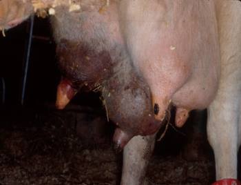
Mastitis is a disease in milch animals. In animals affected by thrush disease, the udder becomes hot in the beginning of the disease and there is pain and swelling in it. Body temperature also increases. The quality of milk is affected as soon as symptoms appear. There is excess of sputum, blood and pus (pus) in milk. The animal stops eating and drinking and becomes disinterested. This disease is commonly found in almost all such animals including cow, buffalo, goat and pig, which feed milk to their young. Tonella disease in animals is caused by infection with a variety of bacteria, viruses, molds and yeasts. Apart from this, due to injury and seasonal adversities, it becomes too dry. Since ancient times, this disease has been a matter of concern for the milking animals and their cattle rearers. This disease alone is the biggest obstacle in the complete success of the White Revolution along with animal wealth development. Crores of rupees are lost every year due to this disease, which ultimately affects the economic condition of livestock farmers.
Symptom
In asymptomatic or asymptomatic type of disease, the udder and milk appear to be quite normal but the disease can be diagnosed by examination of milk in the laboratory. In symptomatic disease, where in some animals there is only pus/splash or blood etc. In some unusual type of disease, the udder rots and falls off. Most of the animals do not have fever etc. If the disease is not treated in time, the general swelling of the udder increases to irreversible and the udder becomes hard like wood. After this stage, the milk coming from the udder stops permanently. Usually one or two udders are affected in the beginning, which can later spread to other udders as well. In some animals the taste of milk changes to salty. The following types of measures can be taken to identify this invisible type of disease in time. 1. Ph. Periodic check of milk by paper or detailed check in case of doubt. 2. Testing through California Mastitis Solution. 3. Milk culture and sensitivity test in case of doubt. Apart from this, proper maintenance of animals, use of medicines to increase immunity of the udder and proper treatment of the disease at all times are preferable.
The treatment
Successful treatment of the disease is possible only in the early stages, otherwise it becomes difficult to save the udder as the disease progresses. To avoid this, the milk of the milch animal should be checked on time and treated with bactericidal drugs by a veterinarian. Often these drugs are given by inserting a tube into the udder as well as by injection into the muscle. The milk of the animal is not drinkable during the treatment by the tube being inserted in the udder. Therefore, milk should not be used until 48 hours after the last tube is mounted. It is very important that the treatment should be done completely, do not leave it in the middle. Furthermore, it should not be expected that (at least) the animal in the present calf will return to normal whole milk after treatment. In order to effectively prevent the disease, it is necessary to pay attention to the following points.
Disease prevention
Anthrax gland disease
Anthrax disease, which is spread in animals through contagion and infection, is fatal. Veterinary experts say that it is an epidemic disease that spreads once in a place, it spreads again and again in the same place. It is also called as Guilty disease, Poisonous fever or Pilbhwa disease. This disease mostly occurs in cow, buffalo, goat and horse. It affects all farm animals. This disease also occurs in humans.
Causes and symptoms of the disease
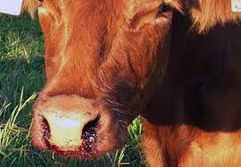
This disease flourishes by eating food and grain contaminated with bacteria, the bacteria of this disease can survive in the soil for 30 years. There is no fixed time or season for the spread of anthrax disease. This disease can spread in any month of the year. This disease is spread by a bacteria. Due to this the animal becomes lethargic and stops gurgling. The sick animal gets high fever. Please enlarge. The stomach becomes very swollen. Bleeding starts from the nose, urinary tract and stool. Sometimes the animal dies suddenly without showing any symptoms.
Protect it like this
According to veterinary experts, it is better to make arrangements for prevention than to treat this disease. It is effective if treated at an early stage, otherwise the animal may die. The animal husbandry must get the disease-preventive vaccine before the disease spreads in the animals, this disease can be prevented by regular annual vaccination in the animals. If vaccinated, the animal is protected from this disease for one year. If anthrax spreads, the animal husbandry should stop the movement of animals to the surrounding village. The buying and selling of skins in the village should be stopped. Do not remove the skin of a dead animal, due to which the disease spreads further. Animals should be buried with lime after digging a pit of five to six feet. One should never open or see the carcass of an animal that has died of Guilty disease. It is transmitted to humans by eating uncooked meat, coming in contact with an infected animal, or inhaling the bacteria. Vaccination should be done at least 1 month before the disease occurs in the particular area.
Version
Anthrax is an infectious disease. Its bacteria live up to 200 years. It spreads through air and water as soon as a favorable environment is found. That's why livestock owners need to be careful with this. When the animal is dead, it should be buried five-six feet deep without post-mortem.
Hemorrhagic Septicemia
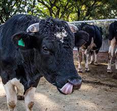
Hemorrhagic septicemia mainly affects cows and buffaloes. This disease is also known by other names like 'Ghurkha', 'Ghontua', 'Ashadhiya', 'Dakaha' etc. The animal becomes a victim of premature death due to this disease. It spreads widely during monsoon. This bacterial disease, which spreads very rapidly, is also contagious. It is caused by a bacterium called Pasteurella multocida. The bacteria of this disease can survive for a long time in moist and moist conditions. This bacterium is present in the upper part of the system in the respiratory tract. Due to the change of weather, the animal becomes vulnerable to the disease. Throat is a very dangerous disease. If the treatment is not started along with the symptoms, the animal dies in a day or two. It has a death rate of more than 80 percent. Begins with high fever (105-107 degrees). A lot of saliva comes out from the mouth of the victim. Swelling in the neck causes wheezing during breathing and eventually death occurs within 12-24 hours. Bury the diseased animal in the pit. If thrown in the open, the infected bacteria spread with water and increase the scope of the disease outbreak.
Symptoms of disease
In this disease the animal suddenly gets high fever and the animal starts trembling. The sick animal becomes lethargic and stops eating and drinking. There is a decrease in milk production. The animal's eyes turn red. The animal is in pain and has difficulty in breathing. There is a wheezing sound in the breath. The animal has a stomach ache, it falls to the ground and saliva also starts pouring out of its mouth. Buffaloes are more sensitive than cows. Animals, especially buffaloes, rarely survive after symptoms appear. Most of the deaths in disease-specific areas occur in older calves and younger adults.
Prevention
Your animals must be vaccinated against this disease every year before the rainy season. All animals of 6 months or more age in a particular area should be tamed before the onset of rains. Avoid gathering of more animals in one place during the rainy season. Sick animals should be kept separate from other healthy animals and separate arrangements should be made for their fodder and water. The place where the animal died should be washed with disinfectant medicines, phenyl or lime solution. Keep the animal house clean and if there is a possibility of disease, immediately contact the veterinarian and seek advice.
The treatment
Treating the animal only when fever starts may save life, otherwise treatment is not effective in this disease. Only a few animals survive once symptoms develop. If left untreated in the early stages of the infection, the mortality rate reaches 100%. Buffalo should be vaccinated deep in the neck meat and after every six months read the information written on it before vaccination. Keep the vaccine at a temperature of two to eight degrees Celsius. The use of antibiotics such as sulfadiamidine oxytetracycline and chloramphoenicol antibiotics are the means of prevention from this disease.